
INTERNATIONAL SOCIETY OF VETERINARY DERMATOPATHOLOGY
ADVANCEMENT OF VETERINARY AND COMPARATIVE DERMATOPATHOLOGY
Welcome to ISVD!
 Login
Login 


Benefits of ISVD Membership



Case of the Month
October 2026
- September 01 - 2023
- Case of the Month
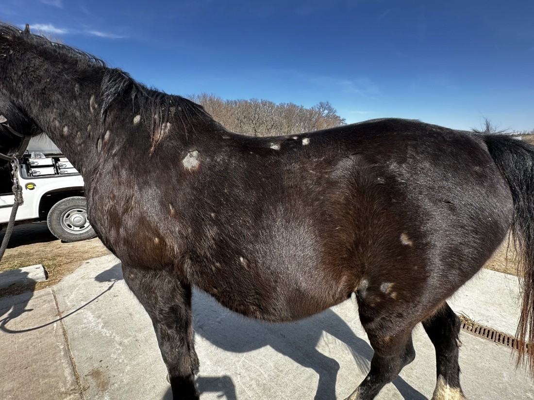
Figure 1. Full body. Case history and Signalment A 19-year-old Arabian gelding from Iowa presented to the ISU Equine Clinic with a progressive hair loss over the last couple of weeks. Physical examination showed moth-eaten alopecia with mild scaling and hyperpigmentation all
Monika Welle Slide Collection

Intro Letter
- October 16 - 2023
- Monika Welle
Dear Colleagues, As some of you might know, I have collected more than 1500 skin slides over the last 25 years, and I would like to make them available to interested veterinary pathologists and veterinary dermatologists, as well as to residents
Veterinary Dermatology
 Veterinary Dermatology is a bi-monthly, peer-reviewed, international journal which publishes papers on all aspects of the skin of mammals, birds, reptiles, amphibians and fish.
Official Journal of ISVD
Members receive a discounted journal subscription with their ISVD membership
> Become a Member or Upgrade
An Amazing Research Tool
VDE publishes original research papers, communications, reviews and letters and will interest researchers, educators and veterinarians in the field of Veterinary Dermatology
Download the App
Keep up with important developments in the field of veterinary dermatology even faster and enjoy an optimized browsing and reading experience on the app
Conferences
Learn of other Veterinary Dermatology events in our profession
|
Figure 1. Full body.
|
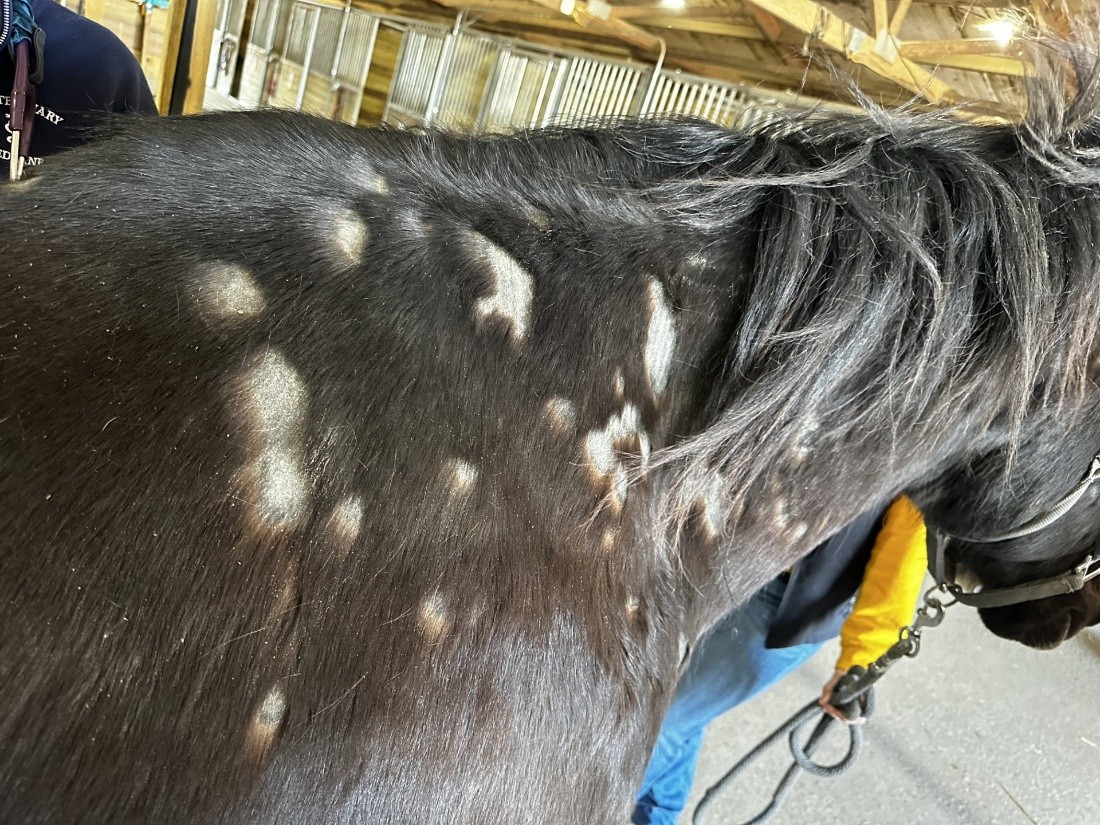
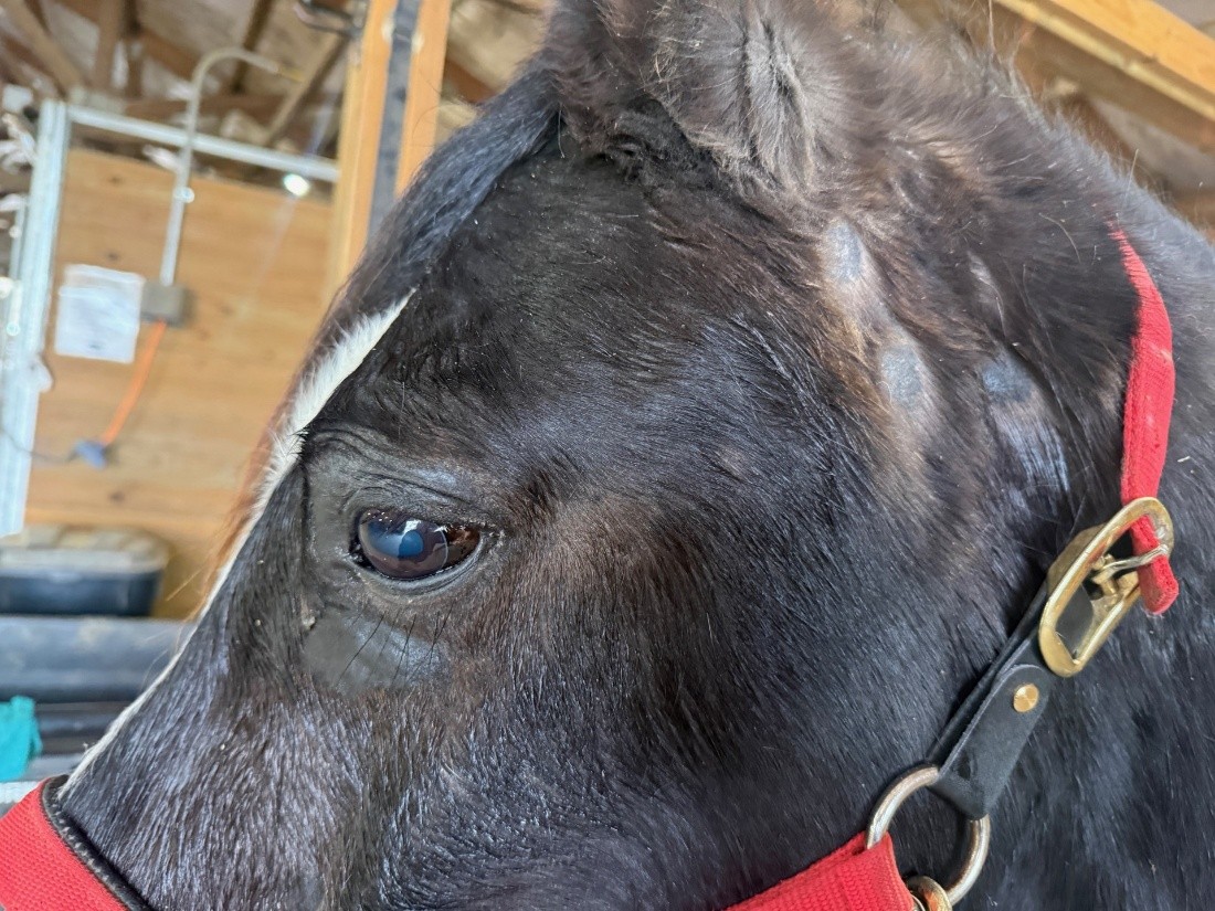
Case history and Signalment A 19-year-old Arabian gelding from Iowa presented to the ISU Equine Clinic with a progressive hair loss over the last couple of weeks. Physical examination showed moth-eaten alopecia with mild scaling and hyperpigmentation all over the body involving the nostrils, periocular, neck, thorax, and appendages. These areas of patchy alopecia range from 0.5-2.5 cm in diameter. The animal showed significant pruritus throughout the examination. Three punch biopsies were taken from the left neck.
|
FIGURES
|
Figure 2: Right side of the neck.
|
|
Figure 3: Left side of the face.
|
|
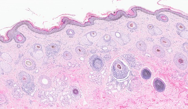
Figure 4: Histopathology. 4X.
|
|
Figure 5: Histopathology. 10X.
|
|
Figure 6: Histopathology. 20X.
|
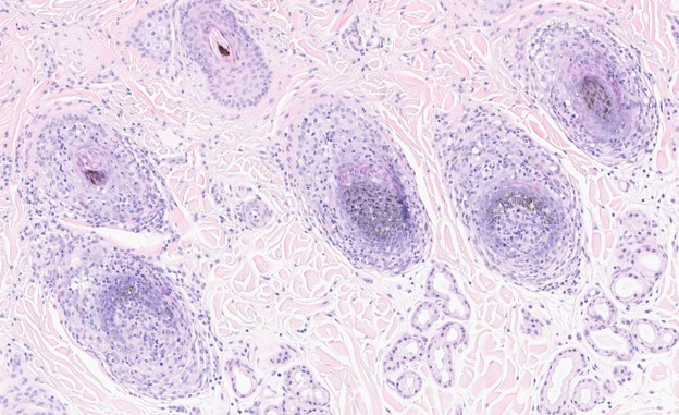
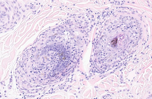
October 2026 - What is the most likely diagnosis of this skin condition?
2025 ISVD Executive Board

Dr. Med Vet, DECVD, MRCVS, RCVS, Assistant Professor Medicine & Epidemiology Department, School of Veterinary Medicine, University of California Davis, USA
DVM, PhD, DECVP, Department of Pathobiology, Texas A&M University, USA

DVM, ECVP, Veterinary Medicine Department, Universita Degli Studi di Perugia, Italy

William Craft DVM, Dipl ACVP, University of Florida, College of Veterinary Medicine, USA

DVM, MS, DACVP, Innovative Vet Path, Leawood, USA

DVM, MS, PhD, DECVD, DACVD, DACVM, University of Florida, College of Veterinary Medicine, USA

DVM, DACVP, Animal Dermatology Group, Inc, Las Vegas, NV, USA

University of California, Davis

Support
|
The ISVD is an affiliate organization of the World Association for Veterinary Dermatology. |
 |