|
Figure 1. |
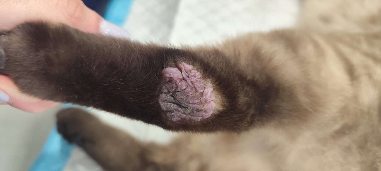
A 1.5-year-old neutered male European Shorthair cat presented with solitary, well-demarcated, alopecic, raised, thin-walled, fluctuant, fluid-filled lesions located over and slightly distal to the hocks. The lesions measured approximately 2–3 cm in diameter, had a verrucous pink surface, and the surrounding haired skin was unremarkable. According to the owner, the lesions had been present for approximately 1–3 months. Additionally, a solitary alopecic, hyperkeratotic, solid plaque measuring approximately 1–2 cm in diameter was present on the dorsal antebrachium. The owner reported frequent licking of all lesions. Wood’s lamp examination was negative, no ectoparasites were identified, and cytologic examination yielded no significant findings. The patient had received corticosteroid therapy within 14 days prior to biopsy collection with minimal clinical improvement. A biopsy specimen from the right hindlimb lesion was submitted for histopathologic examination.
|

FIGURES
|
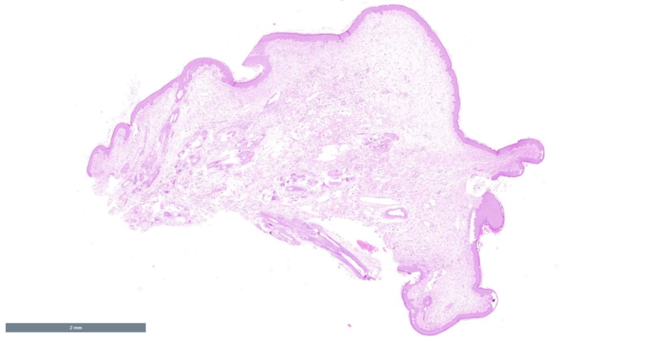
Figure 2. |
Figure 3. H&E. 2x magnification. |
|
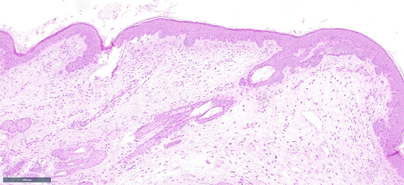
Figure 4. H&E. 10x magnification.
|
|
|
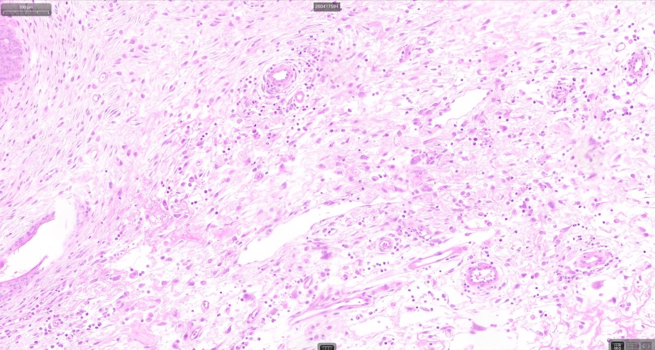
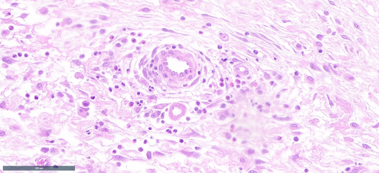
Figure 5. H&E. 40x magnification.
Figure 6. H&E. 40x magnification. |
|